Retinopathy
What is Retinopathy?
Retinopathy is an eye disease that causes the photoreceptor cells in the dog’s retina to degrade over time. More recently retinopathy has been found in the Vallhund breed in the uk, although cases were found in other countries before this time none were confirmed here until 2013. The disease can vary in its severity and onset with some dogs being found affected at around 2-4 years old and other dogs having clear eye tests before being found affected in old age. Retinopathy is not painful and in mild cases the dog may be night blind where it cannot see properly in the dark. In the most severe cases a dog may become blind.
A study into the disease laid out three stages of progression. In stage 1 affected dogs show diffuse multifocal discoloration of the tapetal fundus with no clinical signs of vision loss (age range 1.9 months - 17.8 years).
In Stage 2 retinal degeneration with multifocal, geographic thinning originating at the edges and spreading throughout the tapetal fundus is found, with the majority of dogs reported no vision loss, although some dogs with advanced thinning were reported to experience mild to moderate night blindness (age range 1.1 - 12.6 years).
Stage 3 is marked by diffuse retinal thinning affecting most of the tapetal fundus. These dogs were reported with loss of night vision and impaired day vision, with some having reporting total blindness (age range 9.2 to 15.4 years), (Cooper et al 2014). Rechecking of affected dogs has found that some have not yet progressed past their original diagnosed stage as they have aged and very old dogs in early stages which shows just how variable retinopathy can be.
What causes Retinopathy?
Retinopathy is a hereditary condition. A marker associated with retinopathy was identified by Finnish researchers and a marker test was developed at Genoscoper. While it is unknown if multiple genetic mutations cause retinopathy one mutation that does has been identified by the Animal Health Trust. This mutations mode of inheritance is autosomal recessive and makes the dog very likely to develop retinopathy at some point in its life time, although at what age this would happen it if does is impossible to tell. So for a dog to be affected by this mutation it must inherit a copy of the gene from both parents.
What are breeders doing about Retinopathy?
Originally breeders had to solely rely on pedigree research and eye testing to try to avoid breeding two dogs very likely to produce affected puppies. However the researchers at the Animal Health Trust found a mutation that they believe is a good candidate to cause retinopathy and produced a DNA test for this mutation. Although this mutation does not account for every case of retinopathy in the breed. With the introduction of the DNA test breeders have the option to use this to further help lower the risk of puppies developing retinopathy. Below is information on the tests.
Retinopathy DNA Test: This mutations mode of inheritance is autosomal recessive, so for a dog to be affected by this mutation it must inherit two copies of the gene, one from both parents. With a simple swab from the inside of a dogs cheek the lab can check the dogs DNA for this mutation and report the results back to the owner as either ;
Clear: The dog does not have this mutation and will not develop retinopathy from the tested mutation as the dog does not have it.
Carrier: The dog has one copy of the mutation and as such it will also not develop retinopathy from this tested mutation as it does not have 2 copies. However it can pass this mutation on to its offspring if bred.
Genetically Affected: The dog has two copies of the mutation and as such will be very likely to develop retinopathy at some point in its life time, although when cannot be known. If bred this dog will always pass on one copy of the mutation to its offspring.
However being DNA affected is not a guarantee a dog will definitely develop the condition, there are old dogs who are DNA affected who still have clear eye tests, likewise in the AHT research a small number of dogs who were DNA Carrier/Clear for this mutation were reported as being diagnosed with retinopathy. As such this test is should be used to help guide breeders in their choices rather than as a means to remove dogs from breeding. As such the SVS still recommend eye testing.
Using this test a breeder can make choices when picking who to breed by knowing the possible breeding outcomes.
Clear X Clear = Each puppy born has a 100% chance to be clear, 0% chance to be a carrier or genetically affected.
Clear X Carrier = Each puppy born has a 50/50 chance to be clear or a carrier, 0% chance to be genetically affected.
Clear X Genetically Affected = Each puppy born has a 100% chance to be a carrier, 0% chance of being clear or genetically affected.
Carrier X Carrier = Each puppy born has a 50% chance to be a carrier, 25% chance to be clear and 25% chance of being genetically affected.
Carrier X Genetically Affected = Each puppy born has a 50/50 chance of being born either a carrier or genetically affected.
Genetically Affected X Genetically Affected = Each puppy has a 100% chance to be genetically affected, 0% chance to be clear or a carrier.
The DNA Test
For more information on this test or to order a kit
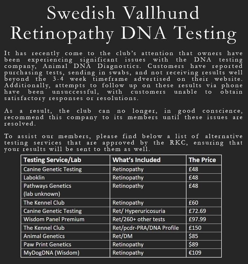
The labs/companies show here are the ones which the kennel club currently recognises the health test results from .
Please check each company to ensure they offer the correct test you require for Swedish Vallhund retinopathy.
(Other companies may also offer the test )
BVA/KC/ISDS Eye Exam: This is an eye exam with certain vets who are on the panel of examiners for the scheme. This exam screens the dogs for eye diseases and will issues a certificate either showing the dog is clear or will list what eye disease the dog has. The exam looks for any eye condition rather than just one specific one. The advice from the scheme is that dogs used for breeding should be re-examined annually .
For more information on this test click here to visit the BVA Website.
What can puppy buyers do?
Your breeder should be happy to show you the result certificates of both mum and dad.
References: Ahonen, S.J., Arumilli, M., Seppälä, E., Hakosalo, O., Kaukonen, M.K.,Komáromy, A.M., Lohi. H (2014). Increased Expression of MERTK is Associated with a Unique Form of Canine Retinopathy. PLoS ONE 9(12): e114552. Cooper, A.E., Ahonen, S., Rowlan, J.S., Duncan, A., Seppälä, E.H., Vanhapelto, P., Lohi, H., Komáromy, A.M (2014). A Novel Form of Progressive Retinal Atrophy in Swedish Vallhund Dogs. PLoS One. 9(9):e106610. Everson, R., Pettitt, L., Forman, O.P., Dower-Tylee, O., McLaughlin, B., Ahonen, S., Kaukonen, M., Komáromy, A.M., Hannes, L., Mellersh, C.S., Sansom, J., Ricketts, S.L. (2017). An Intronic LINE-1 Insertion in MERTK Is Strongly Associated With Retinopathy in Swedish Vallhund Dogs. PLoS One. 12(8):e0183021.